Copyright
©2014 Baishideng Publishing Group Inc.
World J Gastroenterol. Oct 14, 2014; 20(38): 14004-14009
Published online Oct 14, 2014. doi: 10.3748/wjg.v20.i38.14004
Published online Oct 14, 2014. doi: 10.3748/wjg.v20.i38.14004
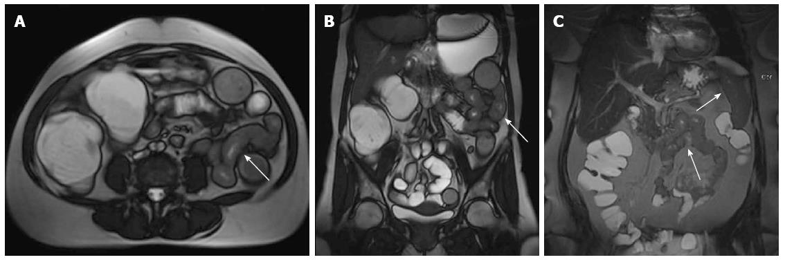
Figure 5 Atypical magnetic resonance enterography findings in a case with small bowel lymphoma.
A: Axial T2 fast imaging with steady-state precession (FISP); B: Coronal T2 FISP; C: T2 maximum intensity projection images. Long segment wall thickening and luminal narrowing in the jejunum (arrows) without accompanying lymphadenopathy. The biopsy showed an isolated small bowel lymphoma, though imaging findings were not strongly suggestive.
- Citation: Cengic I, Tureli D, Aydin H, Bugdayci O, Imeryuz N, Tuney D. Magnetic resonance enterography in refractory iron deficiency anemia: A pictorial overview. World J Gastroenterol 2014; 20(38): 14004-14009
- URL: https://www.wjgnet.com/1007-9327/full/v20/i38/14004.htm
- DOI: https://dx.doi.org/10.3748/wjg.v20.i38.14004


