Copyright
©2014 Baishideng Publishing Group Inc.
World J Gastroenterol. Oct 14, 2014; 20(38): 14004-14009
Published online Oct 14, 2014. doi: 10.3748/wjg.v20.i38.14004
Published online Oct 14, 2014. doi: 10.3748/wjg.v20.i38.14004
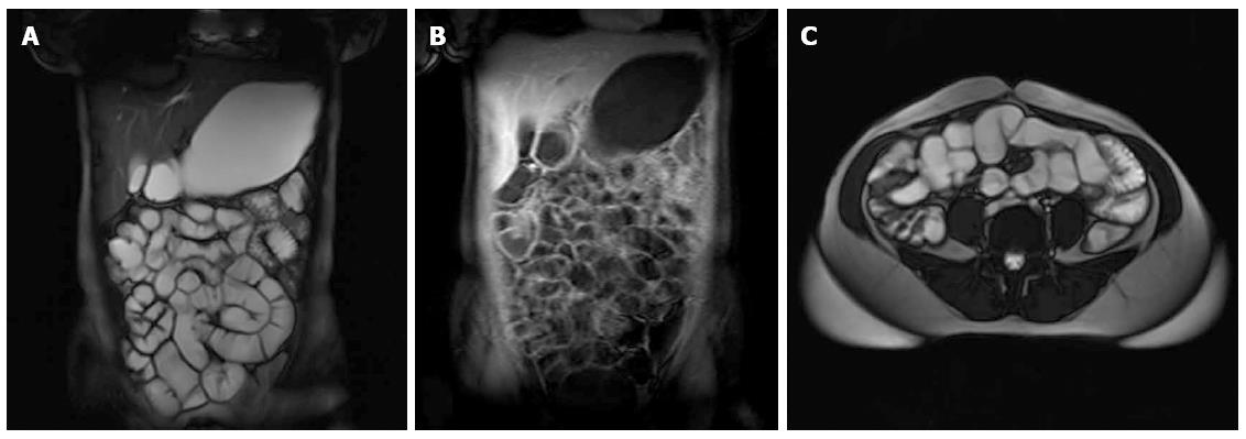
Figure 1 Normal magnetic resonance enterography findings.
A: Coronal T2 fast imaging with steady-state precession (FISP); B: Coronal T1 volumetric interpolated breath-hold examination with contrast; C: Axial T2 FISP images.
- Citation: Cengic I, Tureli D, Aydin H, Bugdayci O, Imeryuz N, Tuney D. Magnetic resonance enterography in refractory iron deficiency anemia: A pictorial overview. World J Gastroenterol 2014; 20(38): 14004-14009
- URL: https://www.wjgnet.com/1007-9327/full/v20/i38/14004.htm
- DOI: https://dx.doi.org/10.3748/wjg.v20.i38.14004


