Copyright
©2014 Baishideng Publishing Group Inc.
World J Gastroenterol. Oct 14, 2014; 20(38): 13842-13862
Published online Oct 14, 2014. doi: 10.3748/wjg.v20.i38.13842
Published online Oct 14, 2014. doi: 10.3748/wjg.v20.i38.13842
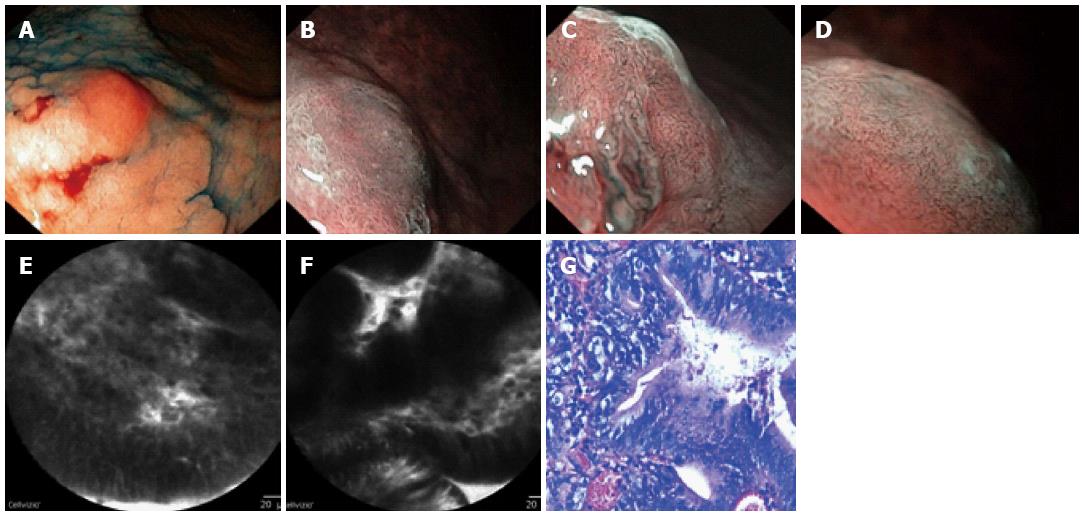
Figure 5 Some patient after 2 wk of Helicobacter pylori eradication therapy.
The characterization of the protruded (Is) part of the lesion type 0-IIa+Is is a differentiated adenocarcinoma. A: High definition Video-EGD + chromoendoscopy: A closer view of the protruded part of the lesion; B-D: Barrow-band imaging (NBI) + zoom: Unclear shredded microsurface pattern with an irregular network microvascular pattern - the capillaries differ in shape and diameter and are tortuous; E: Confocal laser endomicroscopy (CLE): Dark irregular glands; pseudostratified epithelium; irregularly shaped nuclei; F: CLE: Dark irregular glands; pseudostratified epithelium; G: Pathomorphology: A well-differentiated adenocarcinoma.
- Citation: Pasechnikov V, Chukov S, Fedorov E, Kikuste I, Leja M. Gastric cancer: Prevention, screening and early diagnosis. World J Gastroenterol 2014; 20(38): 13842-13862
- URL: https://www.wjgnet.com/1007-9327/full/v20/i38/13842.htm
- DOI: https://dx.doi.org/10.3748/wjg.v20.i38.13842


