Copyright
©2014 Baishideng Publishing Group Inc.
World J Gastroenterol. Oct 7, 2014; 20(37): 13615-13619
Published online Oct 7, 2014. doi: 10.3748/wjg.v20.i37.13615
Published online Oct 7, 2014. doi: 10.3748/wjg.v20.i37.13615
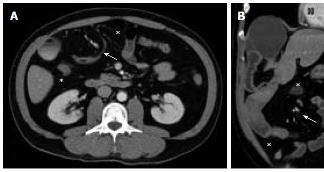
Figure 2 Computed tomography scans showing the incarceration of Meckel’s diverticulum and the small bowel in the right subphrenic space, causing the depression of liver surface.
The twisted mesenteric vessels (arrow) are indicative of a volvulus; the group of small-bowel loops lateral to the colon (asterisk) also suggest a transmesocolic internal hernia. A: Axial view; B: Coronal view.
- Citation: Wu SY, Ho MH, Hsu SD. Meckel's diverticulum incarcerated in a transmesocolic internal hernia. World J Gastroenterol 2014; 20(37): 13615-13619
- URL: https://www.wjgnet.com/1007-9327/full/v20/i37/13615.htm
- DOI: https://dx.doi.org/10.3748/wjg.v20.i37.13615


