Copyright
©2014 Baishideng Publishing Group Inc.
World J Gastroenterol. Oct 7, 2014; 20(37): 13453-13465
Published online Oct 7, 2014. doi: 10.3748/wjg.v20.i37.13453
Published online Oct 7, 2014. doi: 10.3748/wjg.v20.i37.13453
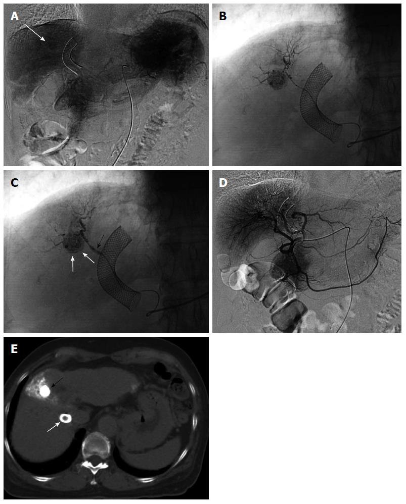
Figure 2 Two-step balloon-occluded transarterial chemoembolization.
A 76-year old woman characterized as Child-Pugh class C underwent transjugular intrahepatic portosystemic shunt (TIPS) for massive ascites before several transarterial chemoembolization (TACE) sessions. Celiac arteriography in the venous phase (A) showed a small hepatocellular carcinoma (HCC) (arrow). A microballoon catheter was advanced into the tumor feeding artery. Then, two-step balloon-TACE (B-TACE) was performed. First, tumors were sufficiently embolized by TACE without balloon occlusion of the tumor feeding artery (B); Second, a microballoon catheter was inflated to occlude the feeding artery (C, black arrow), and B-TACE was performed to fully embolize the peritumoral liver parenchyma (C, white arrows). Common hepatic arteriography showed no proximal migration or leakage of embolization materials (D). Following the two-step B-TACE procedure, computed tomography showed dense lipiodol accumulation in the HCC (E, black arrow). There was no evidence of proximal migration or leakage of embolization materials. White arrows indicated a stent for TIPS.
- Citation: Murata S, Mine T, Sugihara F, Yasui D, Yamaguchi H, Ueda T, Onozawa S, Kumita SI. Interventional treatment for unresectable hepatocellular carcinoma. World J Gastroenterol 2014; 20(37): 13453-13465
- URL: https://www.wjgnet.com/1007-9327/full/v20/i37/13453.htm
- DOI: https://dx.doi.org/10.3748/wjg.v20.i37.13453


