
















©2014 Baishideng Publishing Group Inc.
World J Gastroenterol. Sep 21, 2014; 20(35): 12691-12695
Published online Sep 21, 2014. doi: 10.3748/wjg.v20.i35.12691
Published online Sep 21, 2014. doi: 10.3748/wjg.v20.i35.12691
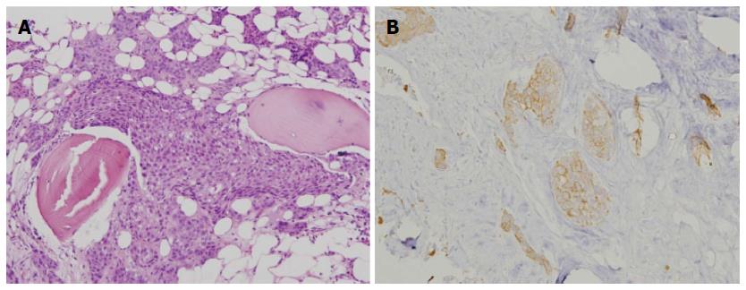
Figure 2 Histologic and immunohistochemical pictures.
A: Histologic picture shows clusters of epithelial-like cells, with eosinophilic cytoplasm and hyperchromatic nuclei. Some nuclei had prominent nucleoli with a high nuclear-to-cytoplasmic ratio. (hematoxylin and eosin staining, original magnification × 100); B: Immunohistochemical picture reveals positivity for high molecular weight (HMW) cytokeratin (original magnification × 200).
- Citation: Chen YH, Huang CH. Esophageal squamous cell carcinoma with dural and bone marrow metastases. World J Gastroenterol 2014; 20(35): 12691-12695
- URL: https://www.wjgnet.com/1007-9327/full/v20/i35/12691.htm
- DOI: https://dx.doi.org/10.3748/wjg.v20.i35.12691