Copyright
©2014 Baishideng Publishing Group Inc.
World J Gastroenterol. Sep 21, 2014; 20(35): 12673-12677
Published online Sep 21, 2014. doi: 10.3748/wjg.v20.i35.12673
Published online Sep 21, 2014. doi: 10.3748/wjg.v20.i35.12673
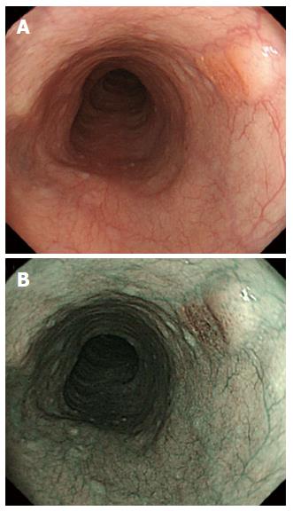
Figure 1 White-light endoscopy revealed a reddish depressed lesion 5 mm in diameter having a subepithelial tumor-like prominence (A), the depressed area was observed as a brownish area by narrowband imaging (B).
- Citation: Kai Y, Kato M, Hayashi Y, Akasaka T, Shinzaki S, Nishida T, Tsujii M, Morii E, Takehara T. Esophageal early basaloid squamous carcinoma with unusual narrowband imaging magnified endoscopy findings. World J Gastroenterol 2014; 20(35): 12673-12677
- URL: https://www.wjgnet.com/1007-9327/full/v20/i35/12673.htm
- DOI: https://dx.doi.org/10.3748/wjg.v20.i35.12673


