Copyright
©2014 Baishideng Publishing Group Inc.
World J Gastroenterol. Aug 28, 2014; 20(32): 11443-11450
Published online Aug 28, 2014. doi: 10.3748/wjg.v20.i32.11443
Published online Aug 28, 2014. doi: 10.3748/wjg.v20.i32.11443
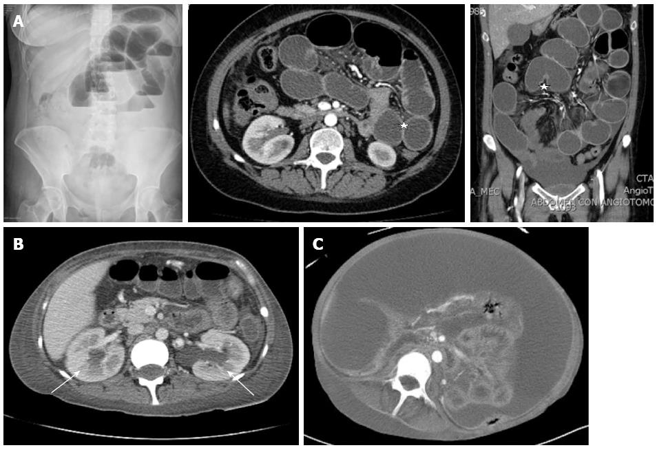
Figure 1 Imaging studies of intestinal pseudo-obstruction in lupus.
A: Represents the images where signs such as distention of the small bowel loops and air fluid levels are visible and edema of the wall labeled with stars known as target lesion; B: Shows the ureterohydronephrosis with labeled arrows (in this case bilateral), which is an accompanying frequent finding; C: Represents the image of intestinal visceromegaly.
- Citation: López CAG, Laredo-Sánchez F, Malagón-Rangel J, Flores-Padilla MG, Nellen-Hummel H. Intestinal pseudo-obstruction in patients with systemic lupus erythematosus: A real diagnostic challenge. World J Gastroenterol 2014; 20(32): 11443-11450
- URL: https://www.wjgnet.com/1007-9327/full/v20/i32/11443.htm
- DOI: https://dx.doi.org/10.3748/wjg.v20.i32.11443


