Copyright
©2014 Baishideng Publishing Group Inc.
World J Gastroenterol. Aug 28, 2014; 20(32): 11080-11094
Published online Aug 28, 2014. doi: 10.3748/wjg.v20.i32.11080
Published online Aug 28, 2014. doi: 10.3748/wjg.v20.i32.11080
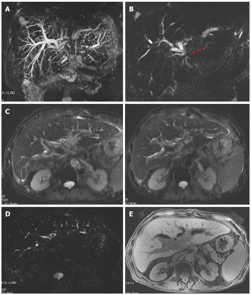
Figure 6 Anastomotic biliary stricture with lithiasis in a patient with hepatico-jejunostomy.
A: Maximum intensity projection reconstruction of 3D thin-slab fast spin-echo T2-weighted images shows marked dilation of the biliary system with partial visualization of the left hepatic duct; B: Single-shot thick-slab magnetic resonance cholangiogram well depicts the stricture of the anastomotic site (red arrow); C, D: Axial single-shot T2-weighted images and axial 3D thin-slab fast spin-echo T2-weighted image demonstrate dilation of the pre-anastomotic biliary tract with the presence of pneumobilia, in particular at the level of left and common hepatic ducts with concomitant stones into the left one; E: Axial T1-weighted image better recognizes pneumobilia.
- Citation: Boraschi P, Donati F. Postoperative biliary adverse events following orthotopic liver transplantation: Assessment with magnetic resonance cholangiography. World J Gastroenterol 2014; 20(32): 11080-11094
- URL: https://www.wjgnet.com/1007-9327/full/v20/i32/11080.htm
- DOI: https://dx.doi.org/10.3748/wjg.v20.i32.11080


