Copyright
©2014 Baishideng Publishing Group Inc.
World J Gastroenterol. Aug 21, 2014; 20(31): 10668-10681
Published online Aug 21, 2014. doi: 10.3748/wjg.v20.i31.10668
Published online Aug 21, 2014. doi: 10.3748/wjg.v20.i31.10668
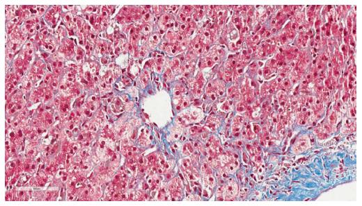
Figure 1 Histopathology of fibrosing cholestatic hepatitis C.
Histopathology of fibrosing cholestatic hepatitis C demonstrating periportal sinusoidal and pericellular “chicken wire” fibrosis (trichrome, image magnification × 40) (Courtesy of Carl Jacobs, MD, Department of Pathology, Carolinas Medical Center, Charlotte, NC, United States).
- Citation: deLemos AS, Schmeltzer PA, Russo MW. Recurrent hepatitis C after liver transplant. World J Gastroenterol 2014; 20(31): 10668-10681
- URL: https://www.wjgnet.com/1007-9327/full/v20/i31/10668.htm
- DOI: https://dx.doi.org/10.3748/wjg.v20.i31.10668


