Copyright
©2014 Baishideng Publishing Group Inc.
World J Gastroenterol. Aug 14, 2014; 20(30): 10440-10448
Published online Aug 14, 2014. doi: 10.3748/wjg.v20.i30.10440
Published online Aug 14, 2014. doi: 10.3748/wjg.v20.i30.10440
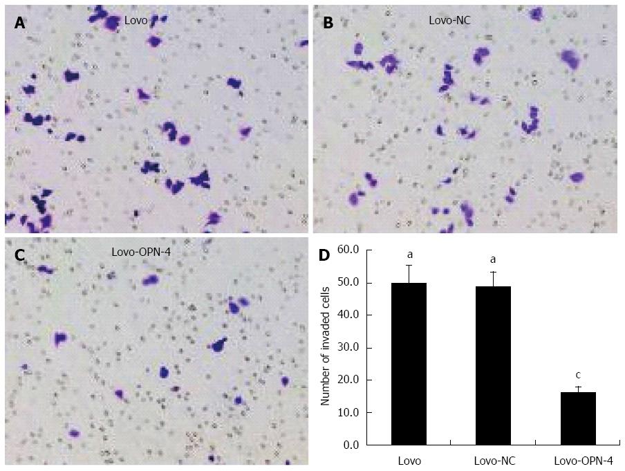
Figure 6 Cell invasion assay.
Staining with 0.1% crystal violet (magnification × 200) revealed a reduced number of invasive Lovo-OPN-4 cells (A-C); Quantification of invasion (D) (n = 15; aP < 0.05 vs Lovo-OPN-4; cP < 0.05 vs Lovo). OPN: Osteopontin.
- Citation: Wu XL, Lin KJ, Bai AP, Wang WX, Meng XK, Su XL, Hou MX, Dong PD, Zhang JJ, Wang ZY, Shi L. Osteopontin knockdown suppresses the growth and angiogenesis of colon cancer cells. World J Gastroenterol 2014; 20(30): 10440-10448
- URL: https://www.wjgnet.com/1007-9327/full/v20/i30/10440.htm
- DOI: https://dx.doi.org/10.3748/wjg.v20.i30.10440


