
















©2014 Baishideng Publishing Group Inc.
World J Gastroenterol. Aug 7, 2014; 20(29): 10202-10207
Published online Aug 7, 2014. doi: 10.3748/wjg.v20.i29.10202
Published online Aug 7, 2014. doi: 10.3748/wjg.v20.i29.10202
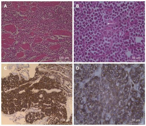
Figure 4 Microscopic examination.
Microscopic examination confirmed that plasma cells had diffusely infiltrated the muscular layer of the stomach (A), and some of these cells contained large nuclei with very prominent centrally located nucleoli (arrow) (B). The immunohistochemical examination disclosed that the tumor cells were positive for CD38 (C) and CD138 (D).
- Citation: Zhao ZH, Yang JF, Wang JD, Wei JG, Liu F, Wang BY. Imaging findings of primary gastric plasmacytoma: A case report. World J Gastroenterol 2014; 20(29): 10202-10207
- URL: https://www.wjgnet.com/1007-9327/full/v20/i29/10202.htm
- DOI: https://dx.doi.org/10.3748/wjg.v20.i29.10202