Copyright
©2014 Baishideng Publishing Group Inc.
World J Gastroenterol. Jul 28, 2014; 20(28): 9497-9505
Published online Jul 28, 2014. doi: 10.3748/wjg.v20.i28.9497
Published online Jul 28, 2014. doi: 10.3748/wjg.v20.i28.9497
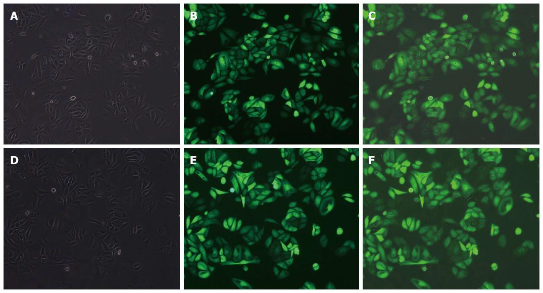
Figure 3 Representative photograph (× 100) showing recombinant adenovirus transfection efficiency evaluated by fluorescence microscopy (transfected with the negative control, top; transfected with the shF1822, bottom).
A, D: Under an ordinary light microscope; B, E: Under a fluorescence microscope; C, F: Superimposed image of the two images.
- Citation: Tao J, Xu XS, Song YZ, Qu K, Wu QF, Wang RT, Meng FD, Wei JC, Dong SB, Zhang YL, Tai MH, Dong YF, Wang L, Liu C. Down-regulation of FoxM1 inhibits viability and invasion of gallbladder carcinoma cells, partially dependent on inducement of cellular senescence. World J Gastroenterol 2014; 20(28): 9497-9505
- URL: https://www.wjgnet.com/1007-9327/full/v20/i28/9497.htm
- DOI: https://dx.doi.org/10.3748/wjg.v20.i28.9497


