Copyright
©2014 Baishideng Publishing Group Inc.
World J Gastroenterol. Jul 14, 2014; 20(26): 8745-8750
Published online Jul 14, 2014. doi: 10.3748/wjg.v20.i26.8745
Published online Jul 14, 2014. doi: 10.3748/wjg.v20.i26.8745
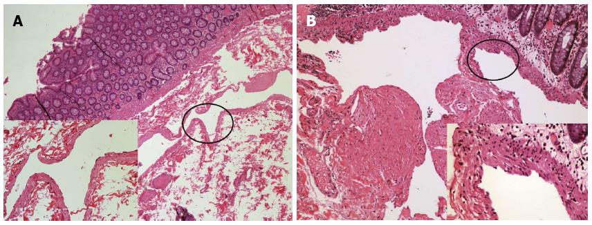
Figure 4 Histopathological examination of the two cases after hematoxylin and eosin stain (at × 40 magnification for low power field images).
A: Case 1 revealed a dilated submucosal cyst lined with a single layer of endothelial cells [left bottom, hematoxylin and eosin (HE) stain, × 200]; B: Case 2 displayed lymphatic vessels of varied sizes, which were covered by a flattened layer of lymphatic epithelium. The smooth muscle layer of the vessels is also noted (right bottom, HE stain, × 400).
- Citation: Zhuo CH, Shi DB, Ying MG, Cheng YF, Wang YW, Zhang WM, Cai SJ, Li XX. Laparoscopic segmental colectomy for colonic lymphangiomas: A definitive, minimally invasive surgical option. World J Gastroenterol 2014; 20(26): 8745-8750
- URL: https://www.wjgnet.com/1007-9327/full/v20/i26/8745.htm
- DOI: https://dx.doi.org/10.3748/wjg.v20.i26.8745


