Copyright
©2014 Baishideng Publishing Group Inc.
World J Gastroenterol. Jul 14, 2014; 20(26): 8745-8750
Published online Jul 14, 2014. doi: 10.3748/wjg.v20.i26.8745
Published online Jul 14, 2014. doi: 10.3748/wjg.v20.i26.8745
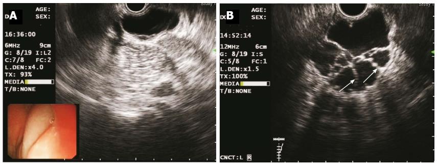
Figure 3 Endoscopic ultrasonography images of the two cases obtained using a catheter EUS probe.
A: Elevated lesions as echo-free, homogenous cysts in Case 1 (frequency 6 MHz); B: Multiple septal walls (indicated by white arrows) in Case 2 (frequency 12 MHz); EUS revealed that both lesions were located in the third (submucosal) layer.
- Citation: Zhuo CH, Shi DB, Ying MG, Cheng YF, Wang YW, Zhang WM, Cai SJ, Li XX. Laparoscopic segmental colectomy for colonic lymphangiomas: A definitive, minimally invasive surgical option. World J Gastroenterol 2014; 20(26): 8745-8750
- URL: https://www.wjgnet.com/1007-9327/full/v20/i26/8745.htm
- DOI: https://dx.doi.org/10.3748/wjg.v20.i26.8745


