Copyright
©2014 Baishideng Publishing Group Inc.
World J Gastroenterol. Jul 14, 2014; 20(26): 8745-8750
Published online Jul 14, 2014. doi: 10.3748/wjg.v20.i26.8745
Published online Jul 14, 2014. doi: 10.3748/wjg.v20.i26.8745
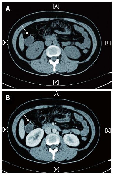
Figure 2 Abdominopelvic computed tomography scan for Case 1 revealed a cystic mass (indicated by white arrow) located at the ascending colon, which was visible in a non-contrast scan (A) and was not enhanced after the arterial phase (B).
A: Anterior; R: Right; L: Left; P: Posterior.
- Citation: Zhuo CH, Shi DB, Ying MG, Cheng YF, Wang YW, Zhang WM, Cai SJ, Li XX. Laparoscopic segmental colectomy for colonic lymphangiomas: A definitive, minimally invasive surgical option. World J Gastroenterol 2014; 20(26): 8745-8750
- URL: https://www.wjgnet.com/1007-9327/full/v20/i26/8745.htm
- DOI: https://dx.doi.org/10.3748/wjg.v20.i26.8745


