Copyright
©2014 Baishideng Publishing Group Inc.
World J Gastroenterol. Jul 7, 2014; 20(25): 8298-8303
Published online Jul 7, 2014. doi: 10.3748/wjg.v20.i25.8298
Published online Jul 7, 2014. doi: 10.3748/wjg.v20.i25.8298
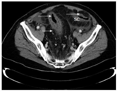
Figure 1 Preoperative computed tomography scan showing edematous sigmoid colon with irregular luminal narrowing within the colonic wall (arrow) and hypoperfusion of the sigmoid colon in the venous phase.
SC: Sigmoid colon.
- Citation: Athanasiou A, Michalinos A, Alexandrou A, Georgopoulos S, Felekouras E. Inferior mesenteric arteriovenous fistula: Case report and world-literature review. World J Gastroenterol 2014; 20(25): 8298-8303
- URL: https://www.wjgnet.com/1007-9327/full/v20/i25/8298.htm
- DOI: https://dx.doi.org/10.3748/wjg.v20.i25.8298


