Copyright
©2014 Baishideng Publishing Group Inc.
World J Gastroenterol. Jun 28, 2014; 20(24): 7752-7759
Published online Jun 28, 2014. doi: 10.3748/wjg.v20.i24.7752
Published online Jun 28, 2014. doi: 10.3748/wjg.v20.i24.7752
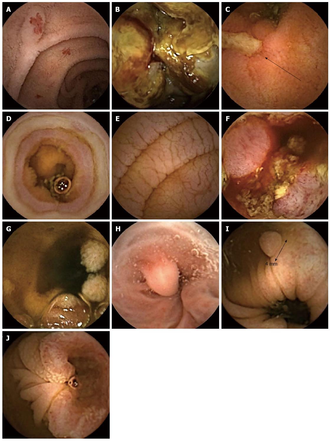
Figure 1 Endoscopy images.
A: Multiple angioectasia; B: Metastatic malignant melanoma of the small bowel; C: Ulceration due to Crohn’s disease-deep ulcer indicated by arrow; D: Circumferential ulceration and stenosis due to non-steroidal anti-inflammatory drug use; E: Typical mucosal changes associated with coeliac disease; F: Enteropathy associated T-cell lymphoma; G: Peutz Jeghers syndrome; H: Oesophageal varix with associated regenerative nodule; I: Colonic polyp; J: Colorectal adenocarcinoma.
- Citation: Hale MF, Sidhu R, McAlindon ME. Capsule endoscopy: Current practice and future directions. World J Gastroenterol 2014; 20(24): 7752-7759
- URL: https://www.wjgnet.com/1007-9327/full/v20/i24/7752.htm
- DOI: https://dx.doi.org/10.3748/wjg.v20.i24.7752


