Copyright
©2014 Baishideng Publishing Group Inc.
World J Gastroenterol. Jun 21, 2014; 20(23): 7381-7391
Published online Jun 21, 2014. doi: 10.3748/wjg.v20.i23.7381
Published online Jun 21, 2014. doi: 10.3748/wjg.v20.i23.7381
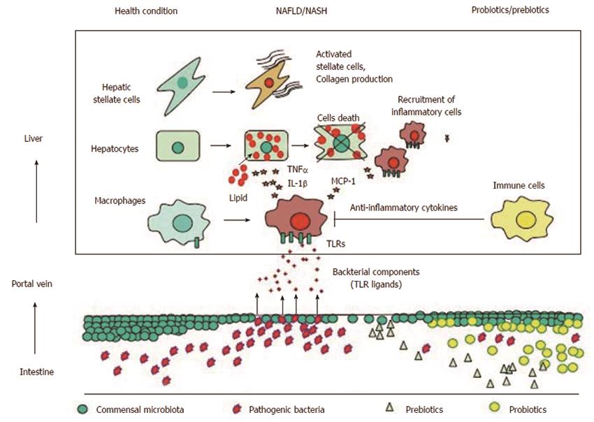
Figure 1 Gut-liver axis in the development of nonalcoholic fatty liver disease.
Under healthy conditions, commensal microbiota inhibit the expansion of pathogenic bacteria and maintain the barrier function of the intestinal epithelium. In nonalcoholic fatty liver disease (NAFLD), the levels of pathogenic bacteria may increase, and the barrier function is disrupted by multiple mechanisms, leading to a translocation of bacteria components [toll-like receptor (TLR) ligands] into the portal vein. TLR ligands stimulate TLR expressing cells, such as macrophages, to produce proinflammatory cytokines including tumor necrosis factor α (TNFα) and interleukin-1b (IL-1b), which promote lipid accumulation as well as hepatocyte cell death. TLR ligands also stimulate macrophages to produce chemokines such as MCP-1, which recruits inflammatory macrophages. These proinflammatory cytokines and certain TLR ligands directly stimulate hepatic stellate cells to produce fibrogenic factors. In contrast, treatments with probiotics or prebiotics protects against the translocation of TLR ligands and the expansion of pathogenic bacteria. In addition, probiotics/prebiotics stimulate immune cells to produce anti-inflammatory cytokines.
- Citation: Miura K, Ohnishi H. Role of gut microbiota and Toll-like receptors in nonalcoholic fatty liver disease. World J Gastroenterol 2014; 20(23): 7381-7391
- URL: https://www.wjgnet.com/1007-9327/full/v20/i23/7381.htm
- DOI: https://dx.doi.org/10.3748/wjg.v20.i23.7381


