Copyright
©2014 Baishideng Publishing Group Inc.
World J Gastroenterol. Jun 21, 2014; 20(23): 7325-7338
Published online Jun 21, 2014. doi: 10.3748/wjg.v20.i23.7325
Published online Jun 21, 2014. doi: 10.3748/wjg.v20.i23.7325
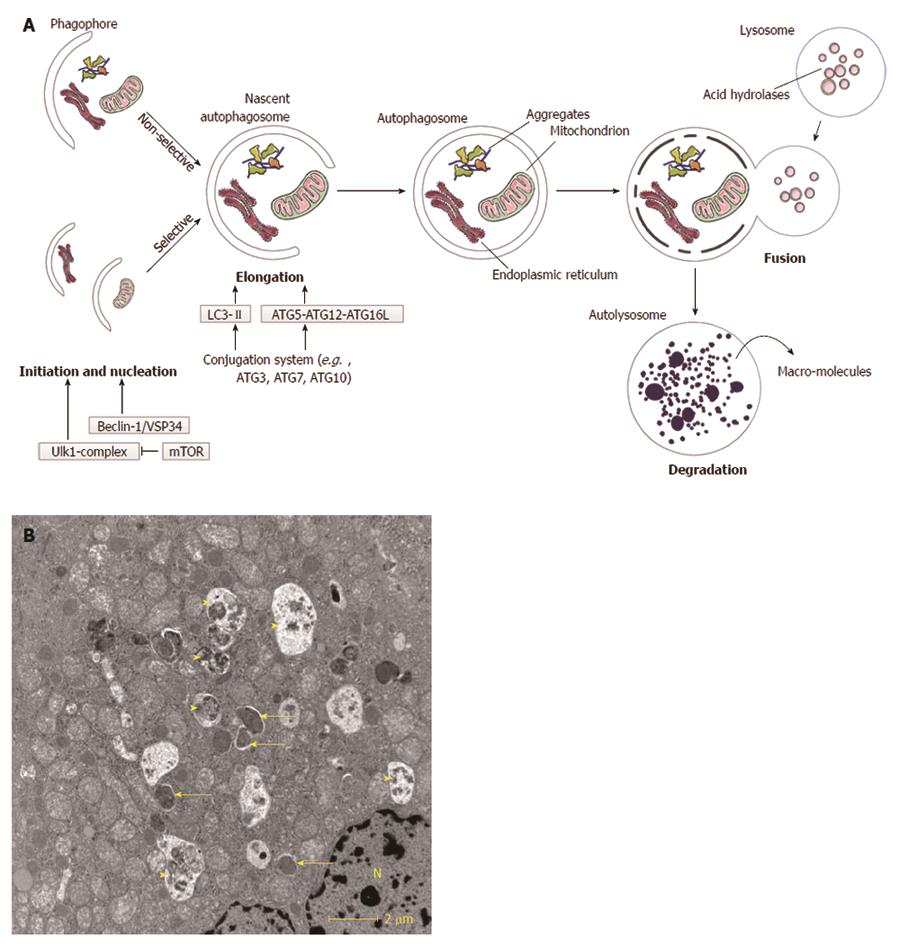
Figure 1 Macroautophagy.
A: Schematic overview of macroautophagy. Macroautophagy starts with the formation of a double-layered membrane, the phagophore (isolation membrane). Phagophore formation is regulated by the ULK1 complex (initiation), which is under control of the mammalian target of rapamycin (mTOR) complex, and the beclin-1/VSP34 interacting complex (nucleation). Two major ubiquitine-like conjugated complexes take care of the elongation of the double membrane: light chain 3 (LC3)-II and ATG5-ATG12-ATG16L1. ATG7 is one of the proteins necessary for formation of both elongation complexes. When an autophagosome is formed, it will fuse with a lysosome. The inner membrane of the autophagosome and the sequestered cytoplasm will be degraded and macromolecules can subsequently be (re-)used. Macroautophagy can be non-selective (random uptake of intracellular material) or selective [uptake of specific cargo, e.g., mitochondria, endoplasmic reticulum (ER)]; B: Transmission electron microsocopy (TEM)-image of a normal mouse liver fasted overnight. The arrows indicate autophagosomes, the arrowheads indicate autolysosomes. N: Nucleus.
- Citation: Kwanten WJ, Martinet W, Michielsen PP, Francque SM. Role of autophagy in the pathophysiology of nonalcoholic fatty liver disease: A controversial issue. World J Gastroenterol 2014; 20(23): 7325-7338
- URL: https://www.wjgnet.com/1007-9327/full/v20/i23/7325.htm
- DOI: https://dx.doi.org/10.3748/wjg.v20.i23.7325


