Copyright
©2014 Baishideng Publishing Group Inc.
World J Gastroenterol. Jun 14, 2014; 20(22): 7075-7078
Published online Jun 14, 2014. doi: 10.3748/wjg.v20.i22.7075
Published online Jun 14, 2014. doi: 10.3748/wjg.v20.i22.7075
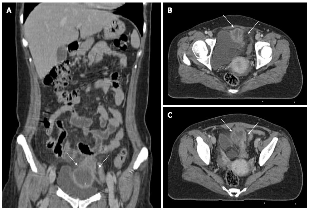
Figure 1 Initial computed tomography image.
A, B: Computed tomography revealed a 3 cm, thin, curvilinear object with a high density within a 5 cm sized fluid collection adjacent to the urinary bladder, which can be regarded as a foreign body and abscess formation (arrow); C: Wall of the rectosigmoid colon (arrow) is thickened, suggesting that the rectosigmoid colon was perforated.
- Citation: Cho MK, Lee MS, Han HY, Woo SH. Fish bone migration to the urinary bladder after rectosigmoid colon perforation. World J Gastroenterol 2014; 20(22): 7075-7078
- URL: https://www.wjgnet.com/1007-9327/full/v20/i22/7075.htm
- DOI: https://dx.doi.org/10.3748/wjg.v20.i22.7075


