Copyright
©2014 Baishideng Publishing Group Inc.
World J Gastroenterol. Jun 7, 2014; 20(21): 6412-6419
Published online Jun 7, 2014. doi: 10.3748/wjg.v20.i21.6412
Published online Jun 7, 2014. doi: 10.3748/wjg.v20.i21.6412
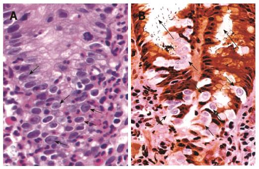
Figure 3 Malgun cell change.
A: Malgun cell change of the epithelial cells at the site of neutrophilic infiltration (arrows), showing large, pale nucleus, prominent nucleolus, and smooth nuclear envelope. They often show characteristic ‘‘perinuclear halo’’ reflecting artificial retraction of cytoplasm (HE, × 150). B: Malgun cells are not stained by silver impregnation in triple silver staining (arrows), demarcating them clearly from other cells. They develop as single cells at the proliferative zone in association with neutrophilic foveolitis, and make small clusters as they move upward to the surface suggesting a clonogenic potential. Note numerous Helicobacter pylori attached to the epithelium and in the lumen (long arrows). (Triple silver staining, × 150).
-
Citation: Lee I. Critical pathogenic steps to high risk
Helicobacter pylori gastritis and gastric carcinogenesis. World J Gastroenterol 2014; 20(21): 6412-6419 - URL: https://www.wjgnet.com/1007-9327/full/v20/i21/6412.htm
- DOI: https://dx.doi.org/10.3748/wjg.v20.i21.6412


