Copyright
©2014 Baishideng Publishing Group Inc.
World J Gastroenterol. May 28, 2014; 20(20): 6180-6200
Published online May 28, 2014. doi: 10.3748/wjg.v20.i20.6180
Published online May 28, 2014. doi: 10.3748/wjg.v20.i20.6180
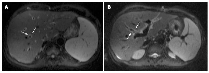
Figure 7 Periportal edema on magnetic resonance diffusion-weighted imaging.
Axial b = 500 s/mm2 diffusion-weighted images acquired on a 54-year-old male patient show a thin rim of hyperintense signal surrounding intrahepatic portal branches (arrows in A and B).
- Citation: Girometti R, Como G, Bazzocchi M, Zuiani C. Post-operative imaging in liver transplantation: State-of-the-art and future perspectives. World J Gastroenterol 2014; 20(20): 6180-6200
- URL: https://www.wjgnet.com/1007-9327/full/v20/i20/6180.htm
- DOI: https://dx.doi.org/10.3748/wjg.v20.i20.6180


