Copyright
©2014 Baishideng Publishing Group Co.
World J Gastroenterol. Jan 14, 2014; 20(2): 598-602
Published online Jan 14, 2014. doi: 10.3748/wjg.v20.i2.598
Published online Jan 14, 2014. doi: 10.3748/wjg.v20.i2.598
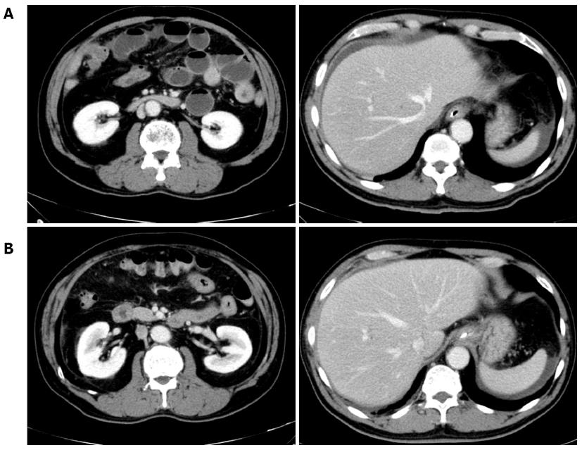
Figure 2 Abdominal computed tomography scan.
A: Showing swelling of the partial segment of the small bowel and dilatation of the intestine (upper left) and ascites around liver and spleen (upper right); B: Demonstrating improvement in the partial swelling of the small bowel and the dilatation of the intestine and ascites collection.
- Citation: Shrestha S, Kisino A, Watanabe M, Itsukaichi H, Hamasuna K, Ohno G, Tsugu A. Intestinal anisakiasis treated successfully with conservative therapy: Importance of clinical diagnosis. World J Gastroenterol 2014; 20(2): 598-602
- URL: https://www.wjgnet.com/1007-9327/full/v20/i2/598.htm
- DOI: https://dx.doi.org/10.3748/wjg.v20.i2.598


