Copyright
©2014 Baishideng Publishing Group Co.
World J Gastroenterol. Jan 14, 2014; 20(2): 486-497
Published online Jan 14, 2014. doi: 10.3748/wjg.v20.i2.486
Published online Jan 14, 2014. doi: 10.3748/wjg.v20.i2.486
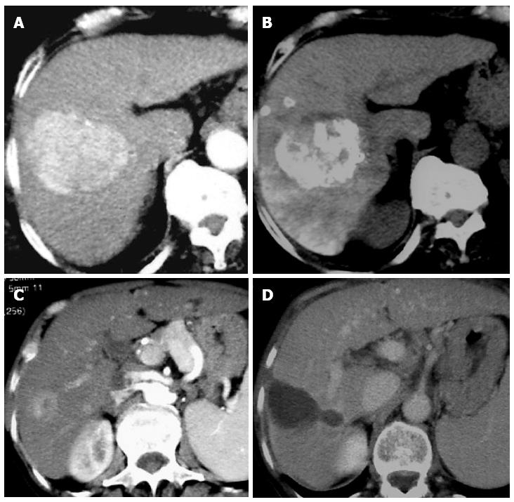
Figure 2 Computed tomography scan.
A: An intermediate hepatocellular cancer (HCC) is showed at liver segment VII before transarterial chemoembolization (TACE). Note the HCC hyperdensity in arterial phase; B: The same tumor showed in A was observed 1 mo after TACE. Note the lipiodol impregnation of the tumor in computed tomography scan without intravenous contrast; C: An early hepatocellular cancer is showed at liver segment V before radiofrequency ablation (RFA). Note the HCC hyperdensity in arterial phase; D: The same tumor showed in C was observed 1 mo after RFA. Note the cavitation of the tumor as an image with no density.
- Citation: Ranieri G, Marech I, Lorusso V, Goffredo V, Paradiso A, Ribatti D, Gadaleta CD. Molecular targeting agents associated with transarterial chemoembolization or radiofrequency ablation in hepatocarcinoma treatment. World J Gastroenterol 2014; 20(2): 486-497
- URL: https://www.wjgnet.com/1007-9327/full/v20/i2/486.htm
- DOI: https://dx.doi.org/10.3748/wjg.v20.i2.486


