Copyright
©2014 Baishideng Publishing Group Co.
World J Gastroenterol. Jan 14, 2014; 20(2): 425-435
Published online Jan 14, 2014. doi: 10.3748/wjg.v20.i2.425
Published online Jan 14, 2014. doi: 10.3748/wjg.v20.i2.425
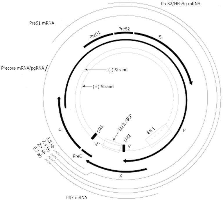
Figure 1 Genomic organization of hepatitis B virus.
The inner circle depicts the rcDNA including the complete minus-strand DNA and the incomplete plus-strand DNA. The direct repeats, DR1 and DR2, as well as the two enhancers, ENI and ENII, are shown. The outer circle depicts the four viral RNAs, the core (C) or pgRNA, the pre-S (L) mRNA, the S mRNA, and the X mRNA. The common 3’-ends (poly-A) of the three mRNAs are indicated by the curve lines. The four protein-coding regions are shown including the precore (PC) and core genes, the polymerase gene, and the X gene. The envelope genes pre-S1 (L), pre-S2 (M), and surface (S) overlap with the polymerase open reading frame.
- Citation: Quarleri J. Core promoter: A critical region where the hepatitis B virus makes decisions. World J Gastroenterol 2014; 20(2): 425-435
- URL: https://www.wjgnet.com/1007-9327/full/v20/i2/425.htm
- DOI: https://dx.doi.org/10.3748/wjg.v20.i2.425


