Copyright
©2014 Baishideng Publishing Group Inc.
World J Gastroenterol. May 21, 2014; 20(19): 5826-5838
Published online May 21, 2014. doi: 10.3748/wjg.v20.i19.5826
Published online May 21, 2014. doi: 10.3748/wjg.v20.i19.5826
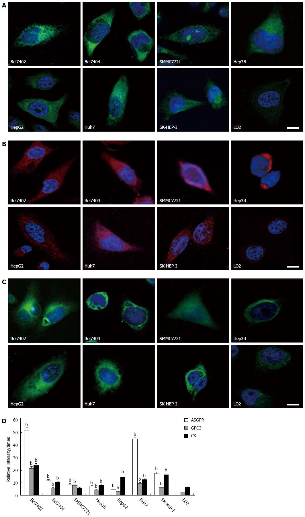
Figure 1 Asialoglycoprotein receptor, glypican-3 and cytokeratin expression, and fluorescence intensities in eight cell lines.
A-C: Representative images of cells on the coverslips with DAPI-stained nuclei (blue) and asialoglycoprotein receptor (ASGPR) staining (green)/glypican-3 (GPC3) staining (red)/cytokeratin (CK) staining (green). The scale bar is 5 μm. D: The bar graph of fluorescence intensities in eight cell lines. Fluorescence intensities were measured using NIH Image J software. Biomarker relative intensities were calculated as the difference between the biomarker staining intensity of the cell and the background intensity. The comparisons between HCC cell lines and the control cell line were analyzed using the Mann-Whitney test (bP < 0.01 vs control).
- Citation: Mu H, Lin KX, Zhao H, Xing S, Li C, Liu F, Lu HZ, Zhang Z, Sun YL, Yan XY, Cai JQ, Zhao XH. Identification of biomarkers for hepatocellular carcinoma by semiquantitative immunocytochemistry. World J Gastroenterol 2014; 20(19): 5826-5838
- URL: https://www.wjgnet.com/1007-9327/full/v20/i19/5826.htm
- DOI: https://dx.doi.org/10.3748/wjg.v20.i19.5826


