Copyright
©2014 Baishideng Publishing Group Co.
World J Gastroenterol. May 14, 2014; 20(18): 5561-5566
Published online May 14, 2014. doi: 10.3748/wjg.v20.i18.5561
Published online May 14, 2014. doi: 10.3748/wjg.v20.i18.5561
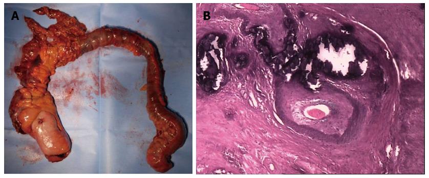
Figure 3 Histological findings.
A: Resected specimen is cyanotic, thick-walled and rigid; B: Microscopic examination demonstrates vascular proliferation in the intestinal sub-mucosa, serosal layer and mesentery. The vascular wall is thickened. Calcification, collagen and hyaline degeneration are detectable.
- Citation: Guo F, Zhou YF, Zhang F, Yuan F, Yuan YZ, Yao WY. Idiopathic mesenteric phlebosclerosis associated with long-term use of medical liquor: Two case reports and literature review. World J Gastroenterol 2014; 20(18): 5561-5566
- URL: https://www.wjgnet.com/1007-9327/full/v20/i18/5561.htm
- DOI: https://dx.doi.org/10.3748/wjg.v20.i18.5561


