Copyright
©2014 Baishideng Publishing Group Co.
World J Gastroenterol. Apr 28, 2014; 20(16): 4546-4557
Published online Apr 28, 2014. doi: 10.3748/wjg.v20.i16.4546
Published online Apr 28, 2014. doi: 10.3748/wjg.v20.i16.4546
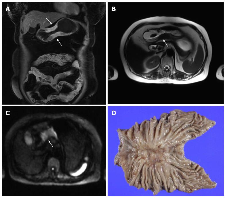
Figure 7 Magnetic resonance imaging of gastric cancer in a 71-year-old female patient.
A: Coronal image of contrast enhanced magnetic resonance imaging (MRI) shows enhancing wall thickening of the gastric body (arrows); B: T2-weighted image delineates a low echoic mass (arrow) destroying layers of the gastric wall. The outer border of the mass is irregular; C: Diffusion-weighted image with a b-value of 1000 shows a high signal intensity mass (arrow) with diffusion restriction; D: Subtotal gastrectomy was performed, and pathological examination revealed pT3 cancer.
- Citation: Choi JI, Joo I, Lee JM. State-of-the-art preoperative staging of gastric cancer by MDCT and magnetic resonance imaging. World J Gastroenterol 2014; 20(16): 4546-4557
- URL: https://www.wjgnet.com/1007-9327/full/v20/i16/4546.htm
- DOI: https://dx.doi.org/10.3748/wjg.v20.i16.4546


