Copyright
©2014 Baishideng Publishing Group Co.
World J Gastroenterol. Apr 28, 2014; 20(16): 4546-4557
Published online Apr 28, 2014. doi: 10.3748/wjg.v20.i16.4546
Published online Apr 28, 2014. doi: 10.3748/wjg.v20.i16.4546
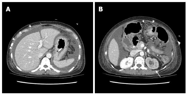
Figure 6 Peritoneal carcinomatosis in a 48-year-old female patient with advanced gastric cancer.
A: Axial 2D CT image shows a large volume of ascites and peritoneal thickening (arrowheads); B: Axial 2D CT image at a lower level than (A) delineates ascites, omental infiltration and nodules (arrowheads), and hydronephrosis of the left kidney (arrow).
- Citation: Choi JI, Joo I, Lee JM. State-of-the-art preoperative staging of gastric cancer by MDCT and magnetic resonance imaging. World J Gastroenterol 2014; 20(16): 4546-4557
- URL: https://www.wjgnet.com/1007-9327/full/v20/i16/4546.htm
- DOI: https://dx.doi.org/10.3748/wjg.v20.i16.4546


