Copyright
©2014 Baishideng Publishing Group Co.
World J Gastroenterol. Apr 28, 2014; 20(16): 4546-4557
Published online Apr 28, 2014. doi: 10.3748/wjg.v20.i16.4546
Published online Apr 28, 2014. doi: 10.3748/wjg.v20.i16.4546
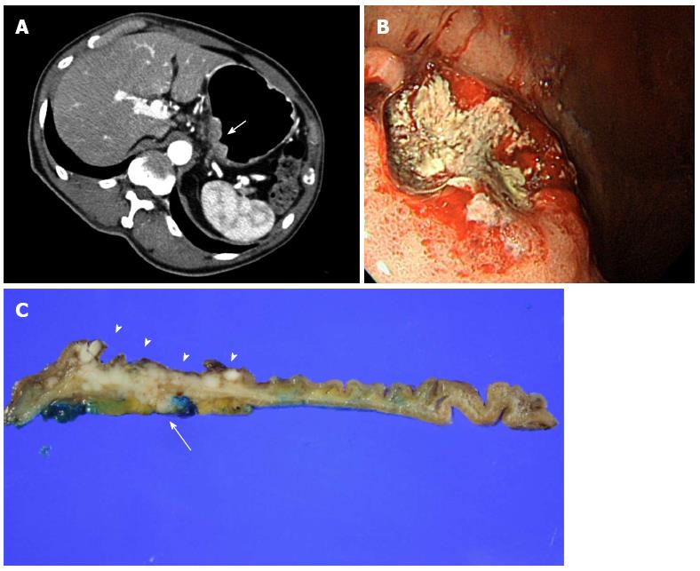
Figure 3 T3 gastric cancer in a 63-year-old male patient.
A: Right decubitus 2D axial image shows thickening of the gastric wall (arrow) involving the entire layer. Perigastric infiltrations are noted outside of the tumor; B: Endoscopy reveals a large ulcerative tumor; C: Surgical specimen showing the tumor (arrowheads) and tumor extension to perigastric fat (arrow).
- Citation: Choi JI, Joo I, Lee JM. State-of-the-art preoperative staging of gastric cancer by MDCT and magnetic resonance imaging. World J Gastroenterol 2014; 20(16): 4546-4557
- URL: https://www.wjgnet.com/1007-9327/full/v20/i16/4546.htm
- DOI: https://dx.doi.org/10.3748/wjg.v20.i16.4546


