Copyright
©2014 Baishideng Publishing Group Co.
World J Gastroenterol. Apr 28, 2014; 20(16): 4546-4557
Published online Apr 28, 2014. doi: 10.3748/wjg.v20.i16.4546
Published online Apr 28, 2014. doi: 10.3748/wjg.v20.i16.4546
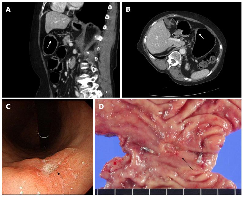
Figure 2 T2 gastric cancer in a 66-year-old female patient.
A: Sagittal 2D image shows enhancing wall thickening with ulceration (arrow) in the lesser curvature side of the low body of the stomach; B: Left posterior oblique axial 2D image also delineates enhancing wall thickening (arrow) in the lesser curvature side of the low body of the stomach. The enhancing lesion involves the entire gastric wall layer, and no low attenuated stripe is visible at the base of the tumor; C: Endoscopy reveals a protruding mass with ulceration (arrow). The impression of the endoscopist was early gastric cancer; D: Subtotal gastrectomy was performed and pathological examination revealed pT2 gastric cancer (arrow).
- Citation: Choi JI, Joo I, Lee JM. State-of-the-art preoperative staging of gastric cancer by MDCT and magnetic resonance imaging. World J Gastroenterol 2014; 20(16): 4546-4557
- URL: https://www.wjgnet.com/1007-9327/full/v20/i16/4546.htm
- DOI: https://dx.doi.org/10.3748/wjg.v20.i16.4546


