Copyright
©2014 Baishideng Publishing Group Co.
World J Gastroenterol. Apr 28, 2014; 20(16): 4546-4557
Published online Apr 28, 2014. doi: 10.3748/wjg.v20.i16.4546
Published online Apr 28, 2014. doi: 10.3748/wjg.v20.i16.4546
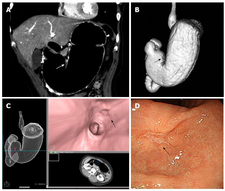
Figure 1 T1a gastric cancer in a 53-year-old female patient.
A: Coronal 2D image shows small mucosal enhancement (arrow) in the lesser curvature side of the antrum; B: Computed tomography gastrography shows a small mucosal irregularity (arrow) in the same area; C: Virtual gastroscopy delineates a shallow, depressed lesion (arrow); D: Endoscopy reveals a small mucosal irregularity confined to the mucosa (arrow). Endoscopic submucosal dissection was performed and pathological examination revealed pT1a early gastric cancer.
- Citation: Choi JI, Joo I, Lee JM. State-of-the-art preoperative staging of gastric cancer by MDCT and magnetic resonance imaging. World J Gastroenterol 2014; 20(16): 4546-4557
- URL: https://www.wjgnet.com/1007-9327/full/v20/i16/4546.htm
- DOI: https://dx.doi.org/10.3748/wjg.v20.i16.4546


