Copyright
©2014 Baishideng Publishing Group Co.
World J Gastroenterol. Apr 21, 2014; 20(15): 4160-4166
Published online Apr 21, 2014. doi: 10.3748/wjg.v20.i15.4160
Published online Apr 21, 2014. doi: 10.3748/wjg.v20.i15.4160
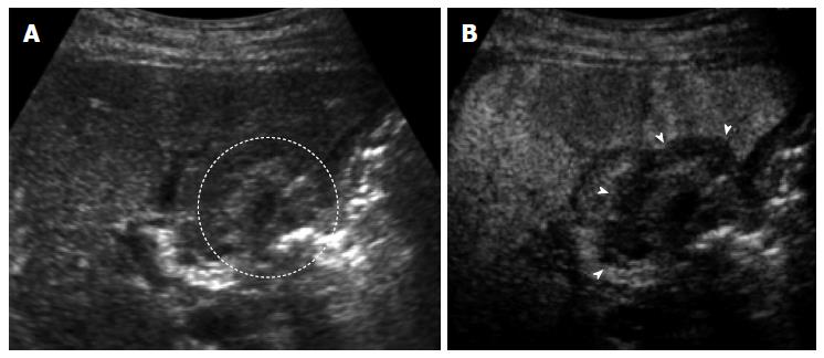
Figure 3 A 70-year-old man with 1.
5 cm hepatocellular carcinoma after radiofrequency ablation. A: The ablated tumor is depicted as hyper echoic lesion (circle) on B-mode ultrasound (US). However, the boundary between ablated area and unablated liver tissue could not be identified clearly; B: Contrast-enhanced US using Sonazoid shows the defect (arrows) in the Kupffer phase. The ablation margin is shown as low echoic zone surrounding the ablated tumor.
- Citation: Minami Y, Nishida N, Kudo M. Therapeutic response assessment of RFA for HCC: Contrast-enhanced US, CT and MRI. World J Gastroenterol 2014; 20(15): 4160-4166
- URL: https://www.wjgnet.com/1007-9327/full/v20/i15/4160.htm
- DOI: https://dx.doi.org/10.3748/wjg.v20.i15.4160


