Copyright
©2014 Baishideng Publishing Group Co.
World J Gastroenterol. Apr 14, 2014; 20(14): 3986-4000
Published online Apr 14, 2014. doi: 10.3748/wjg.v20.i14.3986
Published online Apr 14, 2014. doi: 10.3748/wjg.v20.i14.3986
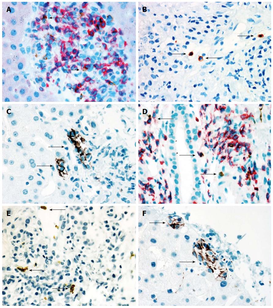
Figure 1 Immunoenzyme stainings in a liver biopsy from a transplanted primary sclerosing cholangitis patient with acute cellular rejection scored according to the Banff criteria as RAI 5 (panels A-C), and from a transplanted control patient (secondary liver metastases) with the same grade of cellular rejection (panels D-F).
Panels A and D show double immunoenzyme staining for CD3 (red) and CD56 (brown, arrows) in a portal area. Panels B and E show immunoperoxidase staining for CD57 (brown, arrows) in a portal area. Panels C and F show CD56-staining of bile ducts (arrows). Only scattered CD56- and CD57 positive leucocytes can been seen in both patients (Original magnification × 400).
- Citation: Fosby B, Næss S, Hov JR, Traherne J, Boberg KM, Trowsdale J, Foss A, Line PD, Franke A, Melum E, Scott H, Karlsen TH. HLA variants related to primary sclerosing cholangitis influence rejection after liver transplantation. World J Gastroenterol 2014; 20(14): 3986-4000
- URL: https://www.wjgnet.com/1007-9327/full/v20/i14/3986.htm
- DOI: https://dx.doi.org/10.3748/wjg.v20.i14.3986


