Copyright
©2014 Baishideng Publishing Group Co.
World J Gastroenterol. Apr 7, 2014; 20(13): 3703-3711
Published online Apr 7, 2014. doi: 10.3748/wjg.v20.i13.3703
Published online Apr 7, 2014. doi: 10.3748/wjg.v20.i13.3703
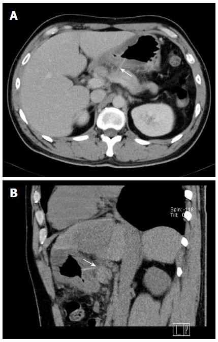
Figure 1 Imaging studies with contrast-enhanced computed tomography scan.
A: A hyperdense linear foreign body embedded in the posterior wall of the antrum with transgastric penetration and attachment to the upper pancreatic border (arrow); B: Sagittal multiplanar reformation image demonstrating close contact of the foreign body to the pancreas without evidence of vascular injury (arrow).
- Citation: Chong LW, Sun CK, Wu CC, Sun CK. Successful treatment of liver abscess secondary to foreign body penetration of the alimentary tract: A case report and literature review. World J Gastroenterol 2014; 20(13): 3703-3711
- URL: https://www.wjgnet.com/1007-9327/full/v20/i13/3703.htm
- DOI: https://dx.doi.org/10.3748/wjg.v20.i13.3703


