Copyright
©2014 Baishideng Publishing Group Co.
World J Gastroenterol. Apr 7, 2014; 20(13): 3698-3702
Published online Apr 7, 2014. doi: 10.3748/wjg.v20.i13.3698
Published online Apr 7, 2014. doi: 10.3748/wjg.v20.i13.3698
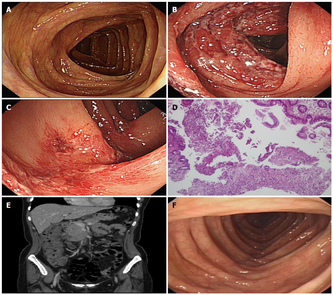
Figure 1 Case 1 findings.
A: Initial colonoscopic findings, Normal colonoscopic findings in ascending colon; B, C Follow up colonoscopic findings; B: The results showed severe petechial hemorrhages with edematous mucosal inflammation in the ascending colon; C: The results showed longitudinal ulcers and hemorrhages in the ascending colon; D: Microscopic findings of affected ascending colon (H and E stain, × 100). Biopsy was compatible with the findings of ischemic colitis, showing erosions and hemorrhages; E: Abdominal computed tomography findings, the results shows diffuse wall thickening of the ascending colon and fluid collection around the ascending colon; F: Follow up colonoscopic findings in 6 mo, the results showed totally normal colonoscopic findings in the ascending colon.
- Citation: Lee SO, Kim SH, Jung SH, Park CW, Lee MJ, Lee JA, Koo HC, Kim A, Han HY, Kang DW. Colonoscopy-induced ischemic colitis in patients without risk factors. World J Gastroenterol 2014; 20(13): 3698-3702
- URL: https://www.wjgnet.com/1007-9327/full/v20/i13/3698.htm
- DOI: https://dx.doi.org/10.3748/wjg.v20.i13.3698


