Copyright
©2014 Baishideng Publishing Group Co.
World J Gastroenterol. Apr 7, 2014; 20(13): 3401-3409
Published online Apr 7, 2014. doi: 10.3748/wjg.v20.i13.3401
Published online Apr 7, 2014. doi: 10.3748/wjg.v20.i13.3401
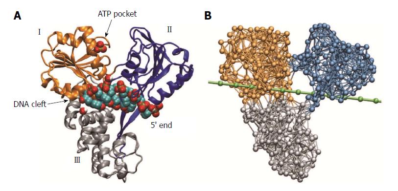
Figure 1 Molecular architecture of hepatitis C virus helicase.
A: Structure of hepatitis C virus (HCV) helicase in ribbon representation (Protein Data Bank code 1A1V). Motor domains I and II are coloured golden and blue, respectively. Domain III is shown in grey. A sulfate ion, shown as beads, is located at the surface of domain I in the proximity of the adenosine-triphosphate (ATP) binding pocket. The short DNA fragment (shown in bead representation) occupies a large cleft separating the motor domains from the third domain; B: Network representation of HCV helicase. The chain of beads representing a single DNA strand is shown in green.
- Citation: Flechsig H. Computational biology approach to uncover hepatitis C virus helicase operation. World J Gastroenterol 2014; 20(13): 3401-3409
- URL: https://www.wjgnet.com/1007-9327/full/v20/i13/3401.htm
- DOI: https://dx.doi.org/10.3748/wjg.v20.i13.3401


