Copyright
©2013 Baishideng Publishing Group Co.
World J Gastroenterol. Mar 7, 2013; 19(9): 1485-1488
Published online Mar 7, 2013. doi: 10.3748/wjg.v19.i9.1485
Published online Mar 7, 2013. doi: 10.3748/wjg.v19.i9.1485
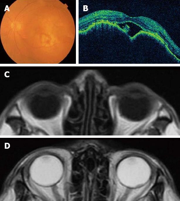
Figure 2 An elevated choroidal neoplasm.
A: Fundoscopic examination; B: Optical coherence tomographic examination; C: Magnetic resonance imaging (MRI) T1-weighted image; D: MRI T2-weighted image.
- Citation: Kawai S, Nishida T, Hayashi Y, Ezaki H, Yamada T, Shinzaki S, Miyazaki M, Nakai K, Yakushijin T, Watabe K, Iijima H, Tsujii M, Nishida K, Takehara T. Choroidal and cutaneous metastasis from gastric adenocarcinoma. World J Gastroenterol 2013; 19(9): 1485-1488
- URL: https://www.wjgnet.com/1007-9327/full/v19/i9/1485.htm
- DOI: https://dx.doi.org/10.3748/wjg.v19.i9.1485


