Copyright
©2013 Baishideng Publishing Group Co.
World J Gastroenterol. Feb 28, 2013; 19(8): 1322-1326
Published online Feb 28, 2013. doi: 10.3748/wjg.v19.i8.1322
Published online Feb 28, 2013. doi: 10.3748/wjg.v19.i8.1322
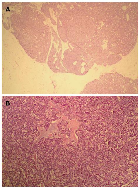
Figure 5 Histological and Pathological analysis of the case (hematoxylin and eosin staining, × 100).
A: Histological analysis shows chief cell hyperplasia of parathyroid gland; B: Pathological analysis shows that tumor cells had acidophilic cytoplasm and round nucleoli which were uniform in size and shape, arranged in tubular, organoid and gyriform patterns.
- Citation: Lu YY, Zhu F, Jing DD, Wu XN, Lu LG, Zhou GQ, Wang XP. Multiple endocrine neoplasia type 1 with upper gastrointestinal hemorrhage and perforation: A case report and review. World J Gastroenterol 2013; 19(8): 1322-1326
- URL: https://www.wjgnet.com/1007-9327/full/v19/i8/1322.htm
- DOI: https://dx.doi.org/10.3748/wjg.v19.i8.1322


