Copyright
©2013 Baishideng Publishing Group Co.
World J Gastroenterol. Feb 28, 2013; 19(8): 1256-1263
Published online Feb 28, 2013. doi: 10.3748/wjg.v19.i8.1256
Published online Feb 28, 2013. doi: 10.3748/wjg.v19.i8.1256
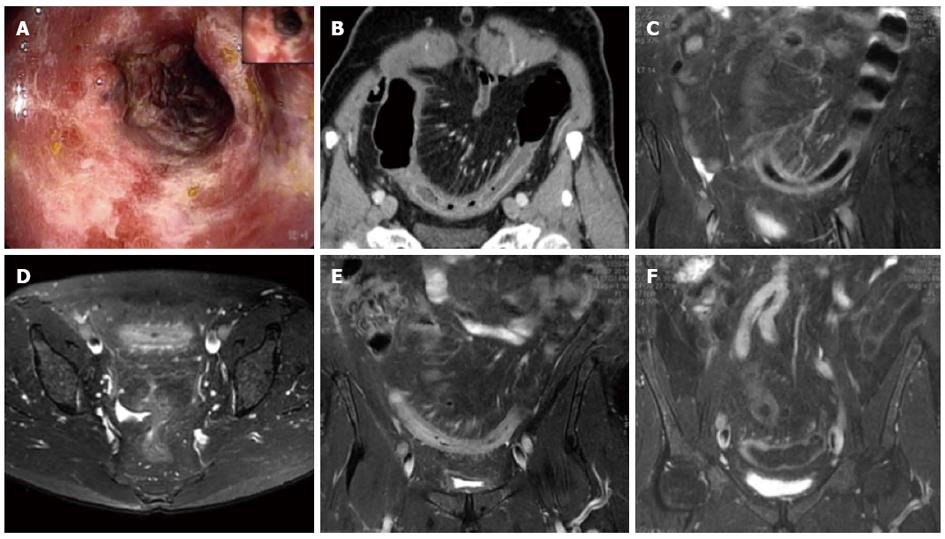
Figure 2 Magnetic resonance imaging follow-up of a patient with ischemic colitis and worsening of clinical symptoms.
Ischemic colitis (IC) of sigmoid colon in a 62-year-old man with left lower quadrant pain and elevate lactate dehydrogenase levels, who presented with melena and a recent history of stenting procedures for ischemic cardiopathy. A: Endoscopic procedure showed multiple necrotic area; B: 2D (two dimensional) coronal reformat contrast-enhanced computed tomography (CT) showed acute IC (Type I CT); C-E: The patient had 2 magnetic resonance examinations (C and D-E) with an interval of 48 h due to worsening of clinical symptoms, with an increase of the length and thickness of the involved tract (D-E); F: The ischemic process resolved without complication after parenteral nutrition, as showed in the follow-up magnetic resonance imaging, performed after 384 h from the date of CT examination.
- Citation: Mazzei MA, Guerrini S, Cioffi Squitieri N, Imbriaco G, Chieca R, Civitelli S, Savelli V, Mazzei FG, Volterrani L. Magnetic resonance imaging: Is there a role in clinical management for acute ischemic colitis? World J Gastroenterol 2013; 19(8): 1256-1263
- URL: https://www.wjgnet.com/1007-9327/full/v19/i8/1256.htm
- DOI: https://dx.doi.org/10.3748/wjg.v19.i8.1256


