Copyright
©2013 Baishideng Publishing Group Co.
World J Gastroenterol. Feb 28, 2013; 19(8): 1256-1263
Published online Feb 28, 2013. doi: 10.3748/wjg.v19.i8.1256
Published online Feb 28, 2013. doi: 10.3748/wjg.v19.i8.1256
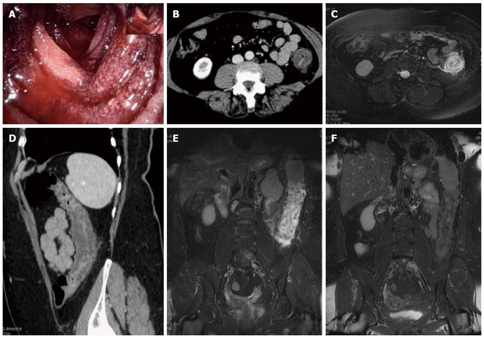
Figure 1 Magnetic resonance imaging follow-up of a patients with ischemic colitis resolved promptly.
Ischemic colitis (IC) of left side colon in a 57 year old woman with a recent history of acute hypertensive crisis, who presented with left lower quadrant pain and massive rectal bleeding. A: Endoscopic procedure showed multiple necrotic area; B and C: Contrast-enhanced computed tomography (CT) and axial T2 fast-recovery fast-spin echo sequence (FRFSE) magnetic resonance imaging (MRI), after 32 h from CT, showed acute IC (Type I CT and MRI) with wall thickening, three layer sandwich sign and a mild amount of free fluid in the paracolic gutter; D and E: 2D coronal reformat CT and coronal T2 FRFSE MRI, at the same time, showed the entire involved tract; F: Ischemia risolved without complications with conservative therapy as shown in the follow-up MRI.
- Citation: Mazzei MA, Guerrini S, Cioffi Squitieri N, Imbriaco G, Chieca R, Civitelli S, Savelli V, Mazzei FG, Volterrani L. Magnetic resonance imaging: Is there a role in clinical management for acute ischemic colitis? World J Gastroenterol 2013; 19(8): 1256-1263
- URL: https://www.wjgnet.com/1007-9327/full/v19/i8/1256.htm
- DOI: https://dx.doi.org/10.3748/wjg.v19.i8.1256


