
















©2013 Baishideng Publishing Group Co.
World J Gastroenterol. Dec 28, 2013; 19(48): 9481-9484
Published online Dec 28, 2013. doi: 10.3748/wjg.v19.i48.9481
Published online Dec 28, 2013. doi: 10.3748/wjg.v19.i48.9481
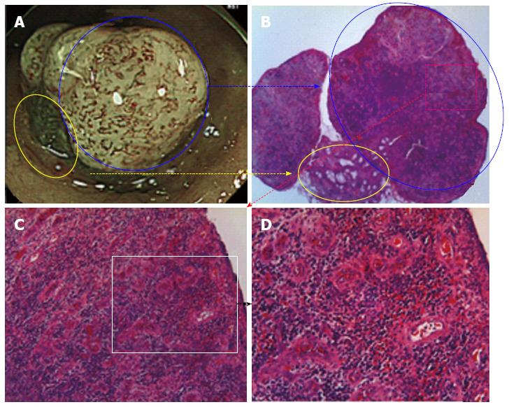
Figure 3 Histological findings of the resected polyp.
A: A 20 magnified narrow band imaging image of the granulomatous polyp; B: A 20 magnified image with a hematoxylin and eosin (HE) stain, the yellow and blue circles in Panel A corresponding to those in Panel B; C: A 100 magnified image with a HE stain reveals significant infiltration of lymphocytes and plasma cells; D: A 200 magnified image with a HE stain reveals increased outgrowth of microvascular structures and infiltration of lymphocytes, neutrophils and plasma cells, which indicates granulation tissue. There were no atypical cells or structural atypia.
- Citation: Mori H, Tsushimi T, Kobara H, Nishiyama N, Fujihara S, Matsunaga T, Ayagi M, Yachida T, Masaki T. Endoscopic management of a rare granulation polyp in a colonic diverticulum. World J Gastroenterol 2013; 19(48): 9481-9484
- URL: https://www.wjgnet.com/1007-9327/full/v19/i48/9481.htm
- DOI: https://dx.doi.org/10.3748/wjg.v19.i48.9481