Copyright
©2013 Baishideng Publishing Group Co.
World J Gastroenterol. Dec 21, 2013; 19(47): 9003-9011
Published online Dec 21, 2013. doi: 10.3748/wjg.v19.i47.9003
Published online Dec 21, 2013. doi: 10.3748/wjg.v19.i47.9003
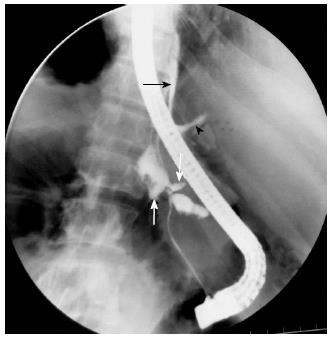
Figure 5 Endoscopic retrograde cholangiopancreatography image.
Another case of traumatic pancreatitis. Fluoroscopic image showing main pancreatic duct disruptions during endoscopic retrograde cholangiopancreatography with multiple contrast filled outpouching is seen, suggestive of pseudocysts (white arrow). Multiple contrast filled tracts are also visualized (black arrowhead). Few tracts are seen in retroperitoneum and one of the tracts is reaching into mediastinum (black arrow). Endoscope is visible.
- Citation: Debi U, Kaur R, Prasad KK, Sinha SK, Sinha A, Singh K. Pancreatic trauma: A concise review. World J Gastroenterol 2013; 19(47): 9003-9011
- URL: https://www.wjgnet.com/1007-9327/full/v19/i47/9003.htm
- DOI: https://dx.doi.org/10.3748/wjg.v19.i47.9003


