Copyright
©2013 Baishideng Publishing Group Co.
World J Gastroenterol. Dec 14, 2013; 19(46): 8722-8730
Published online Dec 14, 2013. doi: 10.3748/wjg.v19.i46.8722
Published online Dec 14, 2013. doi: 10.3748/wjg.v19.i46.8722
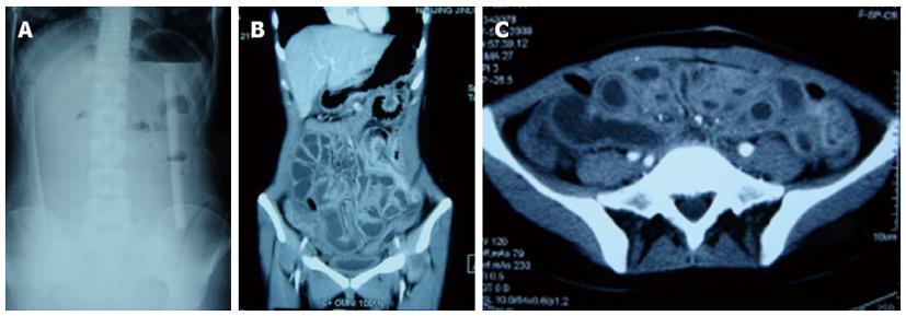
Figure 2 Typical radiographic and intra-operative finding in the last operation.
A: Typical upright radiograph of early postoperative small bowel obstruction with obliterative peritonitis after extensive adhesiolysis for abdominal cocoon, showing only mild air-fluid levels. No isolated small bowel loops were observed; B, C: Computed tomography scan reveals edematous small bowel filled-up with fluids. The border between the small bowel loops is not clear. No significant discrepancies in small bowel diameter and air-fluid levels were observed.
- Citation: Gong JF, Zhu WM, Yu WK, Li N, Li JS. Conservative treatment of early postoperative small bowel obstruction with obliterative peritonitis. World J Gastroenterol 2013; 19(46): 8722-8730
- URL: https://www.wjgnet.com/1007-9327/full/v19/i46/8722.htm
- DOI: https://dx.doi.org/10.3748/wjg.v19.i46.8722


