Copyright
©2013 Baishideng Publishing Group Co.
World J Gastroenterol. Dec 14, 2013; 19(46): 8502-8514
Published online Dec 14, 2013. doi: 10.3748/wjg.v19.i46.8502
Published online Dec 14, 2013. doi: 10.3748/wjg.v19.i46.8502
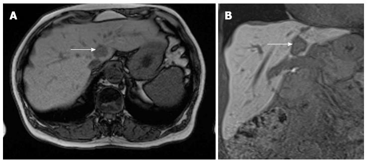
Figure 4 A 63-year-old female with colorectal cancer and suspected liver metastasis.
A: Primovist images acquired 10 min p.i., during the hepatobiliary phase using a T1 VIBE isovoxel sequence with coronal orientation; B: Due to the high resolution axial reconstructions are also done routinely. The lesion in segment I (arrow) is clearly demarcated as a contrast defect because of the missing hepatocytes in the metastasis while the other parts of the liver show a bright contrast enhancement.
- Citation: Kekelidze M, D’Errico L, Pansini M, Tyndall A, Hohmann J. Colorectal cancer: Current imaging methods and future perspectives for the diagnosis, staging and therapeutic response evaluation. World J Gastroenterol 2013; 19(46): 8502-8514
- URL: https://www.wjgnet.com/1007-9327/full/v19/i46/8502.htm
- DOI: https://dx.doi.org/10.3748/wjg.v19.i46.8502


