Copyright
©2013 Baishideng Publishing Group Co.
World J Gastroenterol. Dec 14, 2013; 19(46): 8502-8514
Published online Dec 14, 2013. doi: 10.3748/wjg.v19.i46.8502
Published online Dec 14, 2013. doi: 10.3748/wjg.v19.i46.8502
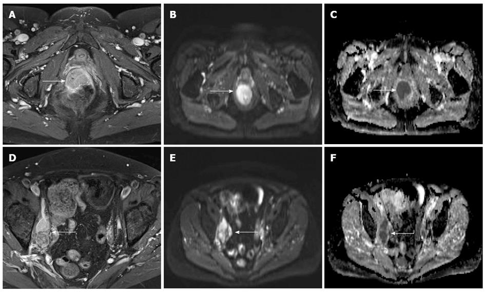
Figure 3 A 58-year-old female with biopsy-proven adenocarcinoma of the rectum.
A: Post-contrast fat-suppressed axial T1 images show a contrast-enhancing mass (arrow), extending from rectum into the anal canal and invading the posterior aspect of the vagina; B, E: Both, the primary tumor and the lymph node metastases, show an hyperintense signal on diffusion weighted imaging; C, F: An reduced apparent diffusion coefficient reflecting the tight tumor cellularity; D: Enlarged, contrast-enhancing lymph nodes along the right iliac axis (arrow).
- Citation: Kekelidze M, D’Errico L, Pansini M, Tyndall A, Hohmann J. Colorectal cancer: Current imaging methods and future perspectives for the diagnosis, staging and therapeutic response evaluation. World J Gastroenterol 2013; 19(46): 8502-8514
- URL: https://www.wjgnet.com/1007-9327/full/v19/i46/8502.htm
- DOI: https://dx.doi.org/10.3748/wjg.v19.i46.8502


