
















©2013 Baishideng Publishing Group Co.
World J Gastroenterol. Dec 7, 2013; 19(45): 8449-8452
Published online Dec 7, 2013. doi: 10.3748/wjg.v19.i45.8449
Published online Dec 7, 2013. doi: 10.3748/wjg.v19.i45.8449
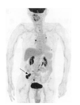
Figure 2 Positron emission tomography and computed tomography showing high metabolism in the right ileum (arrow) and multiple lymph nodes with high metabolism in the mesentery root of the small intestine, in which malignant lesions in the terminal ileum were suspected.
- Citation: Xu XQ, Hong T, Li BL, Liu W. Ileo-ileal intussusception caused by diffuse large B-cell lymphoma of the ileum. World J Gastroenterol 2013; 19(45): 8449-8452
- URL: https://www.wjgnet.com/1007-9327/full/v19/i45/8449.htm
- DOI: https://dx.doi.org/10.3748/wjg.v19.i45.8449