Copyright
©2013 Baishideng Publishing Group Co.
World J Gastroenterol. Nov 28, 2013; 19(44): 8146-8150
Published online Nov 28, 2013. doi: 10.3748/wjg.v19.i44.8146
Published online Nov 28, 2013. doi: 10.3748/wjg.v19.i44.8146
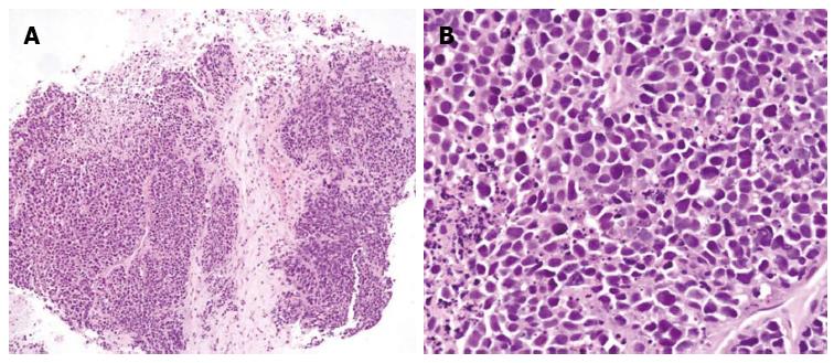
Figure 3 Tumor consisted of tightly packed nests and diffuse, irregularly shaped sheets of cells with necrotic areas.
A: The tumor cells were of small-to-intermediate size with hyperchromatic, round-to-oval nuclei and scanty, poorly defined cytoplasm (HE, × 100); B: The nuclear chromatin was finely granular, and nucleoli were absent or inconspicuous. Cell borders were rarely seen, and nuclear molding was common (HE, × 400).
- Citation: Jo JM, Cho YK, Hyun CL, Han KH, Rhee JY, Kwon JM, Kim WK, Han SH. Small cell carcinoma of the liver and biliary tract without jaundice. World J Gastroenterol 2013; 19(44): 8146-8150
- URL: https://www.wjgnet.com/1007-9327/full/v19/i44/8146.htm
- DOI: https://dx.doi.org/10.3748/wjg.v19.i44.8146


