Copyright
©2013 Baishideng Publishing Group Co.
World J Gastroenterol. Nov 28, 2013; 19(44): 8099-8107
Published online Nov 28, 2013. doi: 10.3748/wjg.v19.i44.8099
Published online Nov 28, 2013. doi: 10.3748/wjg.v19.i44.8099
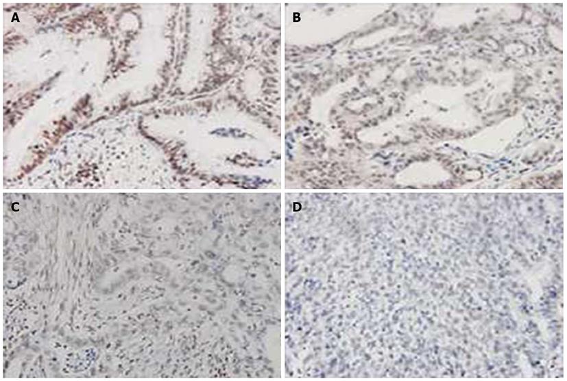
Figure 4 Immunohistochemical detection of uH2B staining at different TNM stages.
A: Stage I (well-differentiated) gastric cancer (GC) tumor shows high-level staining; B: Stage II (moderately-differentiated) GC tumor shows fewer uH2B+ cells and moderate staining (yellow); C: Stage III (poorly-differentiated) GC tumor shows few uH2B+ cells and low-level staining; D: Stage IV (dedifferentiated) GC tumor shows no UH2B+ cells and negative staining. Magnification: × 200.
- Citation: Wang ZJ, Yang JL, Wang YP, Lou JY, Chen J, Liu C, Guo LD. Decreased histone H2B monoubiquitination in malignant gastric carcinoma. World J Gastroenterol 2013; 19(44): 8099-8107
- URL: https://www.wjgnet.com/1007-9327/full/v19/i44/8099.htm
- DOI: https://dx.doi.org/10.3748/wjg.v19.i44.8099


